Home
/ Bone Cross Section Microscope - 3 : Under the stereo microscope (and depending on the section of the bone under investigation) the.
Bone Cross Section Microscope - 3 : Under the stereo microscope (and depending on the section of the bone under investigation) the.
Bone Cross Section Microscope - 3 : Under the stereo microscope (and depending on the section of the bone under investigation) the.. Spongy bone also contains osteocytes housed in lacunae, but they are not arranged in concentric circles. We offer the best quality for the lowest price. Thin section of dinosaur bone. Thin section of dinosaur bone. This model shows a cross section of compact bone.
Osteons (or haversian system) are the structural unit of compact bone. The microscopic cross section measures the probability of occurrence of a particular nuclear reaction. Cross section human tissue microscope viewmedical stock photo 552419425. These are abundant and characteristic of compact bone. This is just a filter on the microscope that disrupts the pathway of light in such a way that you can visualize the orientations of the crystal structure of bone and other hard tissues.
Bone Tissue And Cells Under The Microscope from www.microscopemaster.com There are two ways to study bone histology. Human yellow bone marrow, hematopoietic tissue. Most of the haversian canals in the fossil bone are filled with quartz or calcite. Compact bone is very different from the other tissues you have seen. Hope you enjoy and please. Vintage anatomical drawings of a human head. Neck anatomy and dissection flow cross section at c4. (micrograph provided by the regents of university of michigan.
Get everyday low prices on our wide selection of premium microscopes.
Get everyday low prices on our wide selection of premium microscopes. In three dimensions an osteon is cylindrical in shape. Choose from 1,000s of microscopes. Transverse cross section of compact bone tissue; Human yellow bone marrow, hematopoietic tissue. (micrograph provided by the regents of university of michigan. Slides have to be made this way because the matrix of bone is too hard to be cut with a knife as the other tissues are. (micrograph provided by the regents of university of michigan. In live bone they are open and in thin section would appear dark. There are two ways to study bone histology. Related posts of cross section of human bone diagram. Cross section of a bone diagram. We offer the best quality for the lowest price.
They conduct blood vessels, lymphatics, and nerves throughout the bone. Bone cross section under microscope. Just pick a microscope slide from below and click on it to view under the virtual microscope. Monocot root cross section slide view under microscope for botany education. Before placing your slide on the microscope stage, remember to read the label, examine the slide with your eye and note any visible macroscopic features that might help your examination.
Tissue Biomechanics Bme 615 from silver.neep.wisc.edu Osteons of a compact bone (cross section) under the microscope. Osteons (or haversian system) are the structural unit of compact bone. Observe that the matrix of the bone is deposited in concentric layers that are called lamellae (5). There are two ways to study bone histology. Thus, substantial time is required to count cracks in each cros … In the center of each osteon is the central canal, a space that houses blood vessels and nerves that supply bone. Bone marrow aspiration uses a hollow needle to remove a small sample (about 1 ml) of bone marrow for examination under a microscope. Colobomycter is a very unusual little reptile (actually, it's a parareptile, an extinct reptilian group that was very diverse during the.
Osteons (or haversian system) are the structural unit of compact bone.
The microscopic cross section measures the probability of occurrence of a particular nuclear reaction. Compact bone cross section courtesy: Hope you enjoy and please. Get everyday low prices on our wide selection of premium microscopes. The concept of a nuclear cross section can be quantified physically in terms of characteristic area where a larger area means a larger probability of interaction. This paper addresses the problem of designing experiments to measure microcrack density in cortical bone. The microscopic cross section measures the probability of occurrence of a particular nuclear reaction. There are two ways to study bone histology. Select from premium bone cross section of the highest quality. Bones protect the various organs of the body, produce red and white blood cells, store minerals, provide structure and. Related posts of cross section of human bone diagram human back muscles and bones. Most of the haversian canals in the fossil bone are filled with quartz or calcite. This model shows a cross section of compact bone.
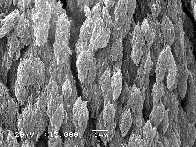
Thus, substantial time is required to count cracks in each cros … Bone under stereo microscope stereo microscopy is one of the simplest methods to view the surface of a bone. This model shows a cross section of compact bone. X75 at 10 cm wide. Conceptual image of human tooth.
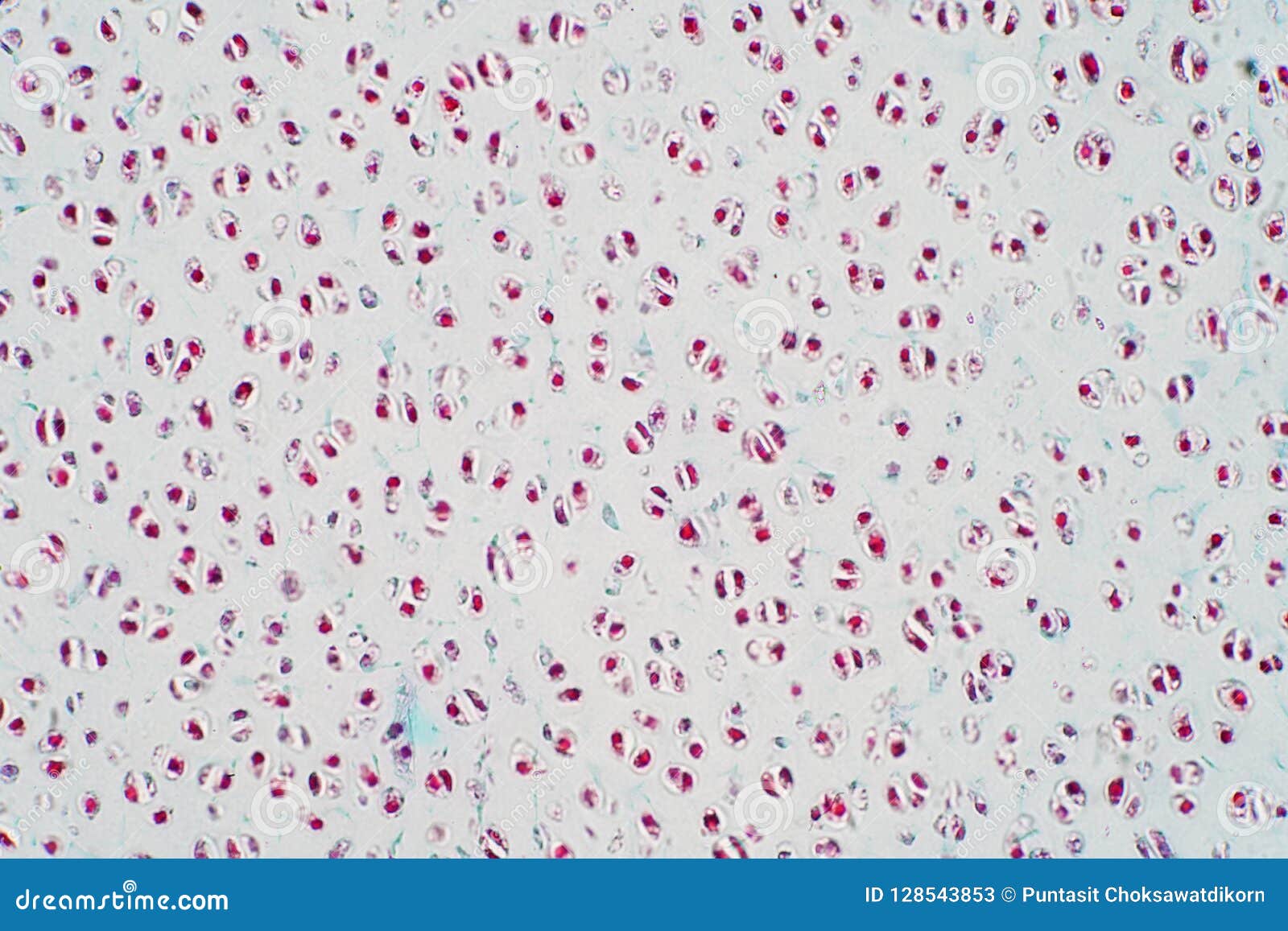
Cross Section Human Cartilage Bone Under Microscope View Stock Image Image Of Fibula Graphic 128543853 from thumbs.dreamstime.com In the center of each osteon is the central canal, a space that houses blood vessels and nerves that supply bone. This paper addresses the problem of designing experiments to measure microcrack density in cortical bone. They are cylindrical structures of concentric layers of bone (lamellae) aligned with the long axis of a bone. Bone cross section illustrations & vectors. Spinal and lumbar disc anatomy. Spongy bone also contains osteocytes housed in lacunae, but they are not arranged in concentric circles. Bone cross section under microscope. Microscope slide showing a cross section of mammalian compact bone.
You can zoom in and explore the different parts of the bone.
When the same lamellar bone is loosely arranged, it is referred to as trabecular bone. Compact bone is very different from the other tissues you have seen. X75 at 10 cm wide. Thin section of dinosaur bone. Bone cross section illustrations & vectors. The microscopic cross section measures the probability of occurrence of a particular nuclear reaction. Select from premium bone cross section of the highest quality. Osteons (or haversian system) are the structural unit of compact bone. We offer the best quality for the lowest price. Thus, substantial time is required to count cracks in each cros … They conduct blood vessels, lymphatics, and nerves throughout the bone. Conceptual image of human tooth. The circular patterns are the concentric lamellae of the haversian canal in the center.
After a fracture, woven bone forms initially and is gradually replaced by lamellar bone during a process known as bony substitution bone cross section. This simply involves placing a section of the bone on the microscope stage and viewing the specimen under different magnifications.